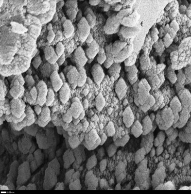
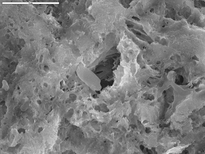
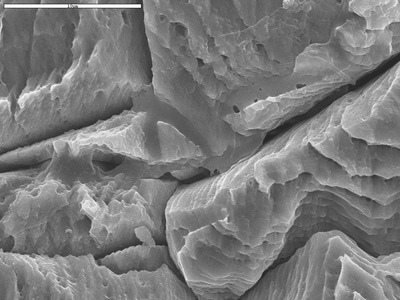
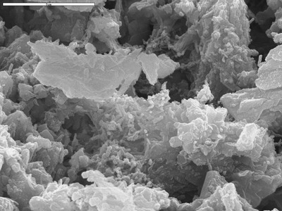
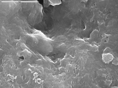
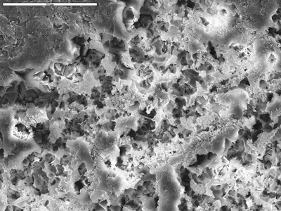
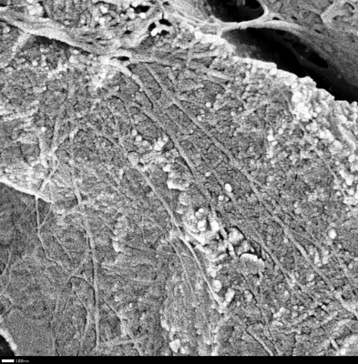
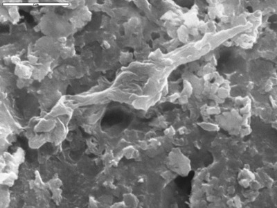
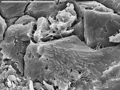
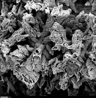
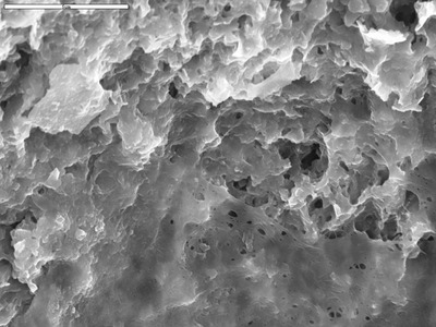
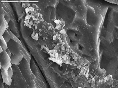
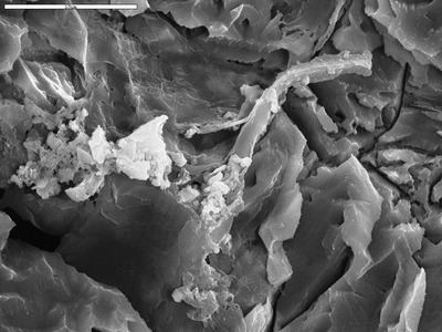
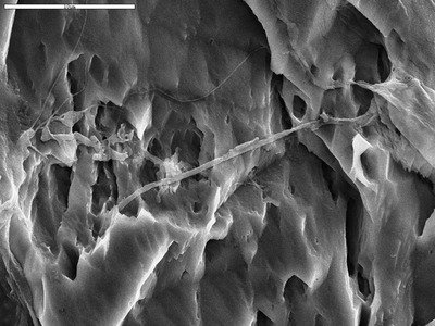
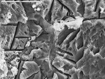
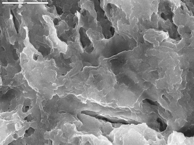
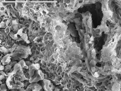
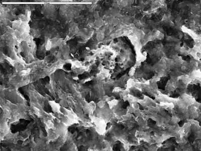
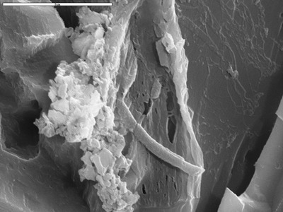
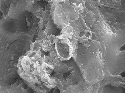
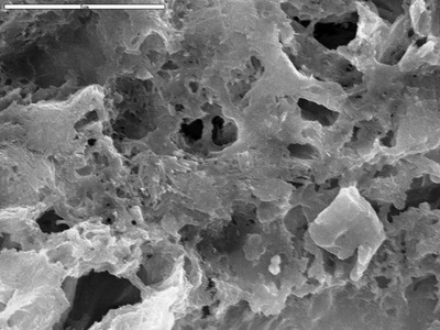
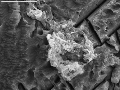
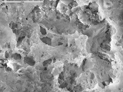
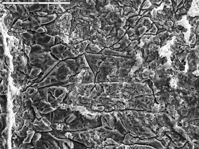
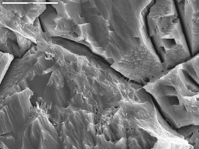
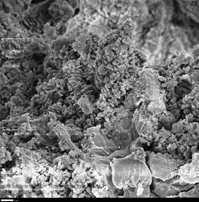
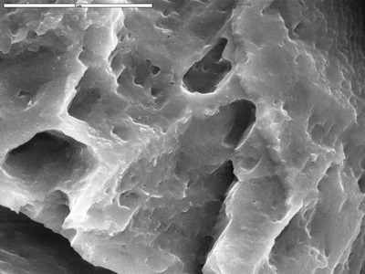
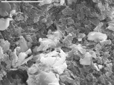
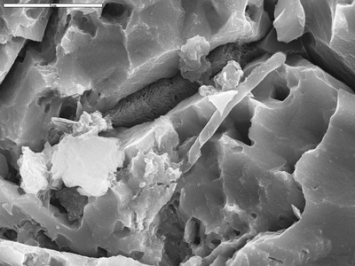
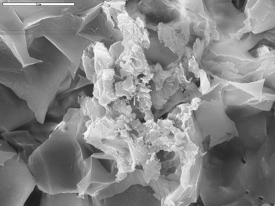
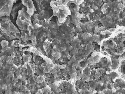
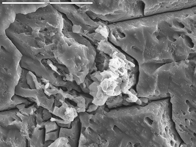
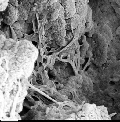
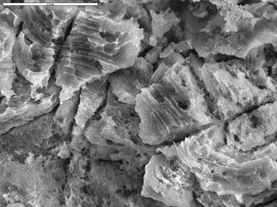
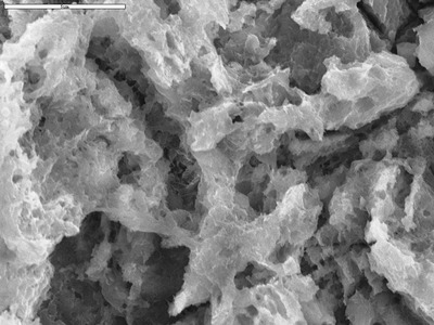
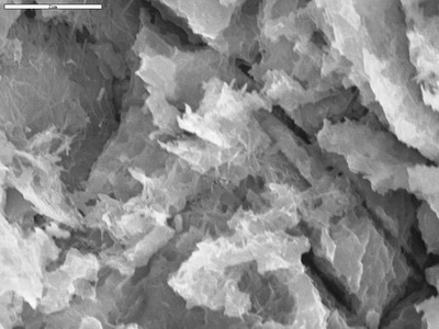
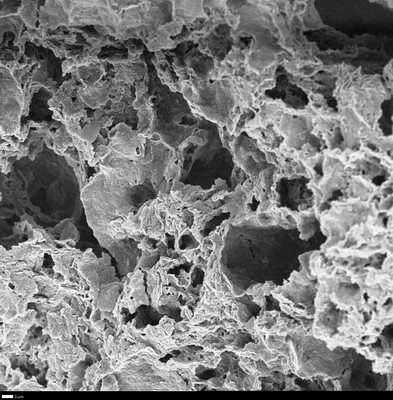
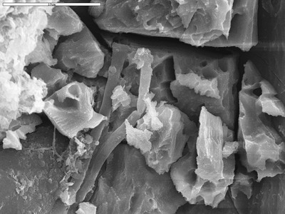
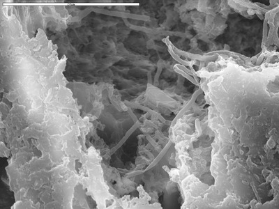
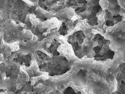
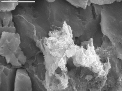
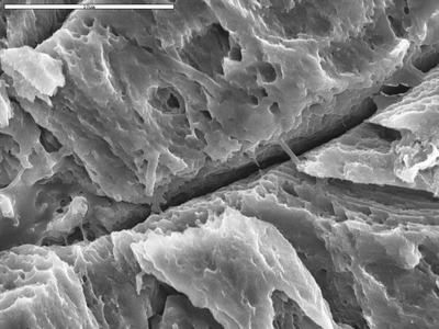
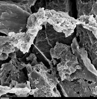
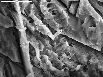
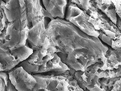
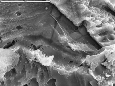
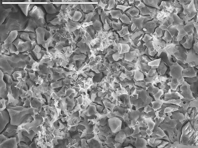
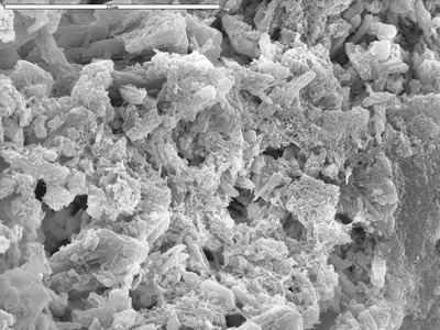
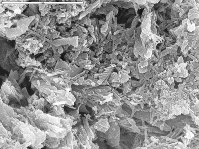
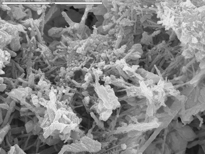
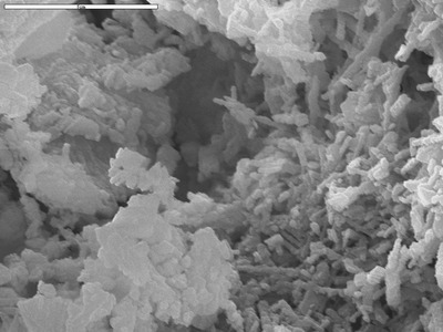
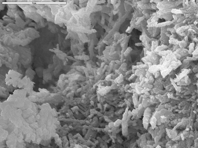
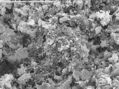
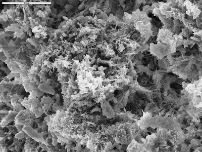
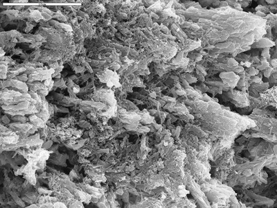
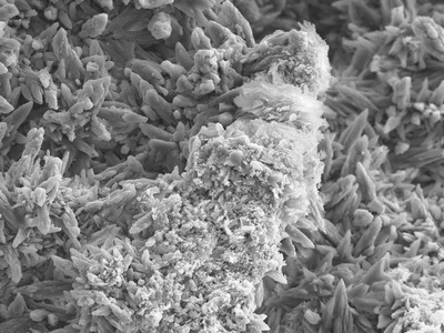
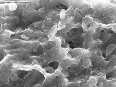
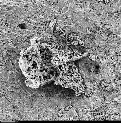
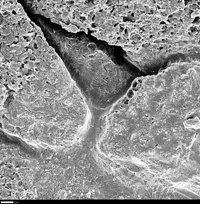
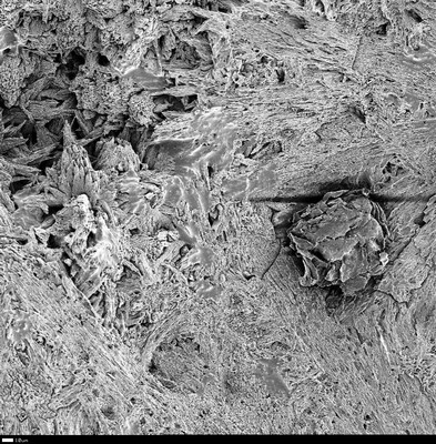
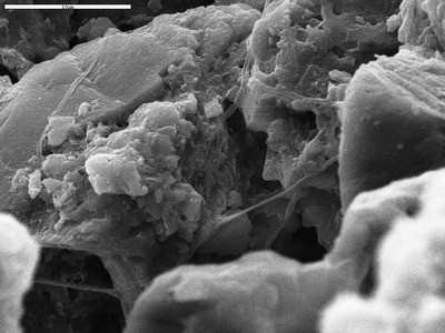
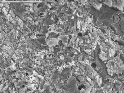
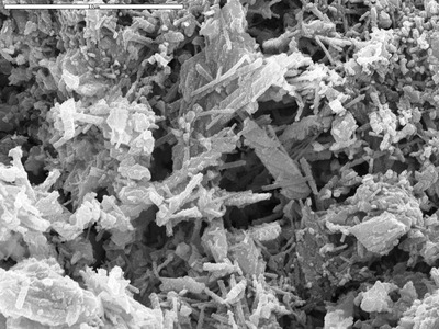
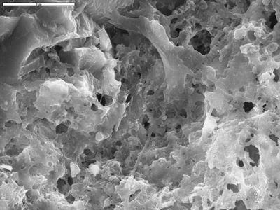
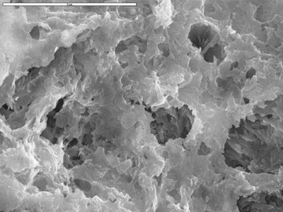
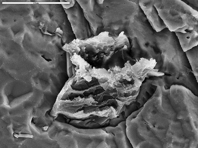
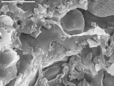
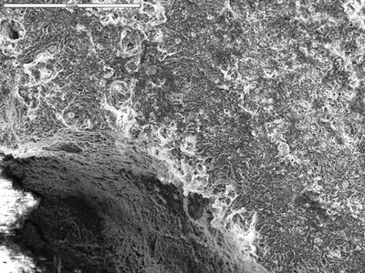
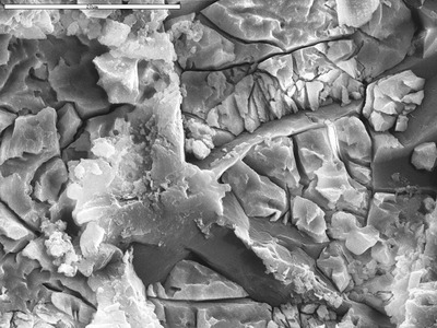
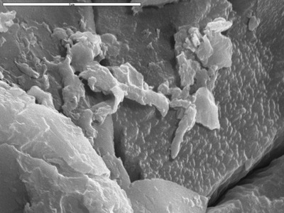
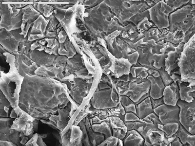
• Home
  • Search
  • Browse Collections
  • My Account
  • About
  • DC Network Digital Commons Network™
Skip to main content
UNM Digital Repository University of New Mexico UNM Digital Repository
  • Home
  • About
  • FAQ
  • My Account

Home > Lib, Mus, & Res Cts > Res Ctrs > Karst and Cave Studies > Pool Precipitates

Pool Precipitates

 
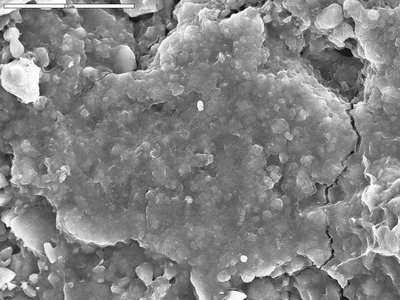
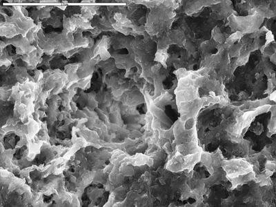
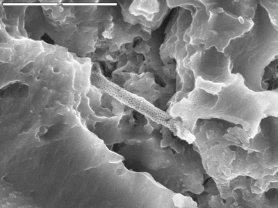
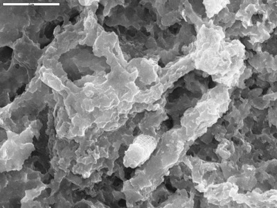
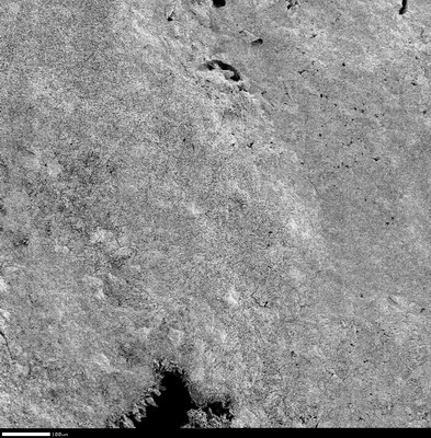
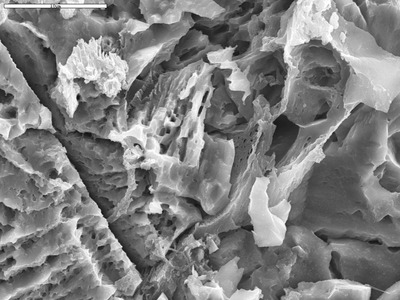
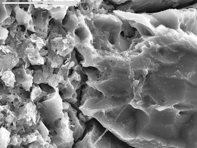
Scanning electron micrographs and spectra. All materials are copyrighted unless otherwise noted.
Printing is not supported at the primary Gallery Thumbnail page. Please first navigate to a specific Image before printing.

Follow

Switch View to List View Slideshow
 
  • Area with many filaments by M. Spilde, D. Northup, and L. Melim

    Area with many filaments

  • Area with no reticulated filaments by D. Northup, M. Splide, L. Melim, R. Liescheidt, and A. Kooser

    Area with no reticulated filaments

  • Area with possible poorly preserved filament by George Braybrook, Leslie Melim, and Brian Jones

    Area with possible poorly preserved filament

  • Area with reticulated filaments, some broken by M. Spilde and Leslie Melim

    Area with reticulated filaments, some broken

  • Area with two smooth filaments and film by D. Northup, M. Spilde, and L. Melim

    Area with two smooth filaments and film

  • A single reticulated filament pressed upon substrate by D. Northup, M. Splide, L. Melim, R. Liescheidt, and A. Kooser

    A single reticulated filament pressed upon substrate

  • Attachment for large filament by M. Spilde and Leslie Melim

    Attachment for large filament

  • Background reading from d50-01 showing Ca by M. Spilde and Leslie Melim

    Background reading from d50-01 showing Ca

  • Backscatter closer view of Mg coating on calcite by M. Spilde, L. Melim, and D. Northup

    Backscatter closer view of Mg coating on calcite

  • Back scatter closer view of odd epoxy string on surface by M. Spilde, L. Melim, and D. Northup

    Back scatter closer view of odd epoxy string on surface

  • Back scatter detail of boundary in Q06-5a-18 by M. Spilde, L. Melim, and D. Northup

    Back scatter detail of boundary in Q06-5a-18

  • Back scatter detail of laminae pinch and recrystallization by M. Spilde, L. Melim, and D. Northup

    Back scatter detail of laminae pinch and recrystallization

  • Back scatter detail of Q06-5a-17, where laminae hit recrystallized patch with epoxy. Bright spots probably Fe grinding grit. by M. Spilde, L. Melim, and D. Northup

    Back scatter detail of Q06-5a-17, where laminae hit recrystallized patch with epoxy. Bright spots probably Fe grinding grit.

  • Back scatter image closer detail of end of laminae in recrystallized patch. by M. Spilde, L. Melim, and D. Northup

    Back scatter image closer detail of end of laminae in recrystallized patch.

  • Back scatter image looking at porous layer within layer 2, laminae go right through the porous material, darker area is epoxy by M. Spilde, L. Melim, and D. Northup

    Back scatter image looking at porous layer within layer 2, laminae go right through the porous material, darker area is epoxy

  • Back scatter image of coarser spar layer that does not cross-cut by M. Spilde, L. Melim, and D. Northup

    Back scatter image of coarser spar layer that does not cross-cut

  • Backscatter image of crystal surface by M. Spilde, D. Northup, and L. Melim

    Backscatter image of crystal surface

  • Back scatter image of detail on boundary to recrystallized area by M. Spilde, L. Melim, and D. Northup

    Back scatter image of detail on boundary to recrystallized area

  • Back scatter image of fine laminae and recrystallization patch with epoxy core by M. Spilde, L. Melim, and D. Northup

    Back scatter image of fine laminae and recrystallization patch with epoxy core

  • Backscatter image of overview of microrods in punky area by M. Spilde, D. Northup, and L. Melim

    Backscatter image of overview of microrods in punky area

  • Back scatter image of Qo6-5a-08, lamina disappears into darker layer with Fe pieces. by M. Spilde, L. Melim, and D. Northup

    Back scatter image of Qo6-5a-08, lamina disappears into darker layer with Fe pieces.

  • Back scatter image of recrystallization area by M. Spilde, L. Melim, and D. Northup

    Back scatter image of recrystallization area

  • Back scatter image overview of C film with Fe spects, and epoxy-filled crack by M. Spilde, L. Melim, and D. Northup

    Back scatter image overview of C film with Fe spects, and epoxy-filled crack

  • Back scatter image showing Fe grain and film and laminae by M. Spilde, L. Melim, and D. Northup

    Back scatter image showing Fe grain and film and laminae

  • Back scatter of clay and calcite mixture by D. Northup, M. Spilde, L. Melim, and R. Liescheidt

    Back scatter of clay and calcite mixture

  • Back scatter of clay and calcite mixture by D. Northup, M. Spilde, L. Melim, and R. Liescheidt

    Back scatter of clay and calcite mixture

  • Backscatter of crust by M. Spilde, D. Northup, and L. Melim

    Backscatter of crust

  • Backscatter of overview of globular grains, and possible clays by M. Spilde, D. Northup, and L. Melim

    Backscatter of overview of globular grains, and possible clays

  • Back scatter of the edge of detrital core, laminae and recrystallization patches, including dolomite rhombs by M. Spilde, L. Melim, and D. Northup

    Back scatter of the edge of detrital core, laminae and recrystallization patches, including dolomite rhombs

  • Back scatter on etched bands by D. Northup, M. Spilde, L. Melim, and R. Liescheidt

    Back scatter on etched bands

  • Back scatter overview of another area with cross-cut laminae and recrystallization by M. Spilde, L. Melim, and D. Northup

    Back scatter overview of another area with cross-cut laminae and recrystallization

  • Back scatter overview of crust on detrital before lamina by M. Spilde, L. Melim, and D. Northup

    Back scatter overview of crust on detrital before lamina

  • Back scatter overview of laminae and recrystallization patterns by M. Spilde, L. Melim, and D. Northup

    Back scatter overview of laminae and recrystallization patterns

  • Backscatter overview showing circular feature with darker ring by M. Spilde, D. Northup, and L. Melim

    Backscatter overview showing circular feature with darker ring

  • Badly etched reticulated filament by M. Spilde, D. Northup, and L. Melim

    Badly etched reticulated filament

  • Barite patch by M. Spilde, D. Northup, and L. Melim

    Barite patch

  • Barite patch in backscatter by M. Spilde, D. Northup, and L. Melim

    Barite patch in backscatter

  • Base of euhedral dogtooth crystals by George Braybrook, Leslie Melim, and Brian Jones

    Base of euhedral dogtooth crystals

  • Biofilm and biofilm threads from pf2p-14 by Michael Spilde and Leslie Melim

    Biofilm and biofilm threads from pf2p-14

  • Biofilm in spar layer by D. Northup, M. Spilde, and L. Melim

    Biofilm in spar layer

  • Bladed crystals with irregular coating by M. Spilde, D. Northup, and L. Melim

    Bladed crystals with irregular coating

  • Boundary between dark layer (upper left) and light layer (lower right) by M. Spilde

    Boundary between dark layer (upper left) and light layer (lower right)

  • Bridging filament by D. Northup and Leslie Melim

    Bridging filament

  • Bridging filament by M. Spilde

    Bridging filament

  • Bridging filament and film by D. Northup, M. Splide, L. Melim, R. Liescheidt, and A. Kooser

    Bridging filament and film

  • Bridging filament, possible reticualted by D. Northup, M. Splide, L. Melim, R. Liescheidt, and A. Kooser

    Bridging filament, possible reticualted

  • Bridging filaments by George Braybrook, Leslie Melim, and Brian Jones

    Bridging filaments

  • Bridging film by George Braybrook, Leslie Melim, and Brian Jones

    Bridging film

  • Bridging film by M. Spilde, D. Northup, and L. Melim

    Bridging film

  • Bridging film and filaments by M. Spilde and L. Melim

    Bridging film and filaments

  • Bridging film and round bodies by George Braybrook, Leslie Melim, and Brian Jones

    Bridging film and round bodies

  • Bridging film in clay by D. Northup, M. Spilde, and L. Melim

    Bridging film in clay

  • Bridging film in coarse crystals area by D. Northup and L. Melim

    Bridging film in coarse crystals area

  • Bridging film with clays by M. Spilde, D. Northup, L. Melim, and R. Liescheidt

    Bridging film with clays

  • Bridging reticulated filaments by M. Spilde, D. Northup, and L. Melim

    Bridging reticulated filaments

  • Bridging ropy film by D. Northup and L. Melim

    Bridging ropy film

  • Bridging smooth filament in film by M. Spilde

    Bridging smooth filament in film

  • Broken crusts revealing inside of formations by George Braybrook, Leslie Melim, and Brian Jones

    Broken crusts revealing inside of formations

  • Broken filament coming out of hole by M. Spilde, D. Northup, L. Melim, and R. Liescheidt

    Broken filament coming out of hole

  • Broken filaments by George Braybrook, Leslie Melim, and Brian Jones

    Broken filaments

  • Broken reticulated filament by George Braybrook, Leslie Melim, and Brian Jones

    Broken reticulated filament

  • Broken reticulated filament bridging surface by M. Spilde and L. Melim

    Broken reticulated filament bridging surface

  • Broken reticulated filament overview by M. Spilde, D. Northup, L. Melim, and R. Liescheidt

    Broken reticulated filament overview

  • Broken reticulated filament pieces by George Braybrook, Leslie Melim, and Brian Jones

    Broken reticulated filament pieces

  • BSE image of round feature from tp1-02.tif by M. Spilde, L. Melim, and D. Northup

    BSE image of round feature from tp1-02.tif

  • BSE overview of white flakey layer by M. Spilde and Leslie Melim

    BSE overview of white flakey layer

  • Calcified film and filaments by M. Spilde and Leslie Melim

    Calcified film and filaments

  • Calcite crystals and epoxy by M. Spilde, L. Melim, and D. Northup

    Calcite crystals and epoxy

  • Calcite growing around more calcite by M. Spilde and Leslie Melim

    Calcite growing around more calcite

  • Center section of reticulated filament in crystals by M. Spilde and Leslie Melim

    Center section of reticulated filament in crystals

  • Clay and calcite mixture by D. Northup, M. Spilde, L. Melim, and R. Liescheidt

    Clay and calcite mixture

  • Clay and film with smooth filament by Michael Spilde

    Clay and film with smooth filament

  • Clay area in lower part of image with etched calcite crystals above by M. Spilde and Leslie Melim

    Clay area in lower part of image with etched calcite crystals above

  • Clay around hole by Michael Spilde

    Clay around hole

  • Clay rich film by D. Northup, M. Splide, L. Melim, R. Liescheidt, and A. Kooser

    Clay rich film

  • Clays and/or filaments etched out of sample by M. Spilde, D. Northup, and L. Melim

    Clays and/or filaments etched out of sample

  • Clays coating calcite by M. Spilde, D. Northup, and L. Melim

    Clays coating calcite

  • Clays on calcite by M. Spilde, D. Northup, and L. Melim

    Clays on calcite

  • Clays standing up within area of epxoy by M. Spilde, D. Northup, and L. Melim

    Clays standing up within area of epxoy

  • Close detail of smooth and hairy filaments by George Braybrook, Leslie Melim, and Brian Jones

    Close detail of smooth and hairy filaments

  • Close detail on spore-like body by George Braybrook, Leslie Melim, and Brian Jones

    Close detail on spore-like body

  • Closer detail of end of laminae in recrystallized patch. by M. Spilde, L. Melim, and D. Northup

    Closer detail of end of laminae in recrystallized patch.

  • Closer detail of lamina by M. Spilde, L. Melim, and D. Northup

    Closer detail of lamina

  • Closer detail of reticulated filament by M. Spilde, L. Melim, S. Herpin, and J.M. Queen

    Closer detail of reticulated filament

  • Closer detail of reticulated filament by M. Spilde, L. Melim, S. Herpin, and J.M. Queen

    Closer detail of reticulated filament

  • Closer of crystals with crust by M. Spilde, L. Melim, and D. Northup

    Closer of crystals with crust

  • Closer overview of biofilm area from d50-20 by M. Spilde and Leslie Melim

    Closer overview of biofilm area from d50-20

  • Closer overview of crystallites by George Braybrook, Leslie Melim, and Brian Jones

    Closer overview of crystallites

  • Closer shot of line of dark S-rich spots by M. Spilde, D. Northup, and L. Melim

    Closer shot of line of dark S-rich spots

  • Closer up of resistant gypsum spots showing imprint of a bladed shape. by M. Spilde and L. Melim

    Closer up of resistant gypsum spots showing imprint of a bladed shape.

  • Closer view, etched interlocking crystals by George Braybrook, Leslie Melim, and Brian Jones

    Closer view, etched interlocking crystals

  • Closer view of aligned crystals by George Braybrook, Leslie Melim, and Brian Jones

    Closer view of aligned crystals

  • Closer view of area with high Mn by M. Spilde, D. Northup, and L. Melim

    Closer view of area with high Mn

  • Closer view of broken reticulated filament by M. Spilde

    Closer view of broken reticulated filament

  • Closer view of coarser, wider reticulated filament by M. Spilde and Leslie Melim

    Closer view of coarser, wider reticulated filament

  • Closer view of contact between dense and punk layers by George Braybrook, Leslie Melim, and Brian Jones

    Closer view of contact between dense and punk layers

  • Closer view of crystal ends and coating by M. Spilde, L. Melim, and D. Northup

    Closer view of crystal ends and coating

  • Closer view of crystal ends and coating by M. Spilde, L. Melim, and D. Northup

    Closer view of crystal ends and coating

  • Closer view of crystallites that look like they coat microrods by George Braybrook, Leslie Melim, and Brian Jones

    Closer view of crystallites that look like they coat microrods

  • Closer view of crystals covered with aligned crystallites by George Braybrook, Leslie Melim, and Brian Jones

    Closer view of crystals covered with aligned crystallites


Page 2 of 16

  • 1
  • 2
  • 3
  • 4
  • 5
  • 6
  • 7


Search

Advanced Search

  • Notify me via email or RSS

Browse

  • Collections
  • Disciplines
  • Authors

Author Corner

  • Author FAQ

Gallery Locations

  • View gallery on map
  • View gallery in Google Earth
 
Elsevier - Digital Commons

Home | About | FAQ | My Account | Accessibility Statement

Privacy Copyright