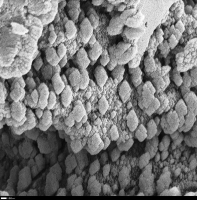
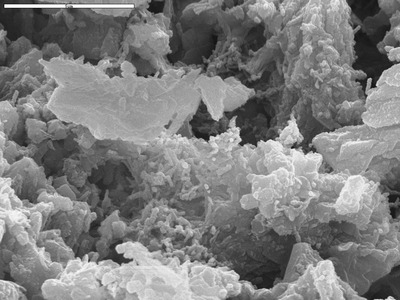
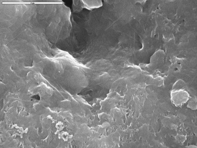
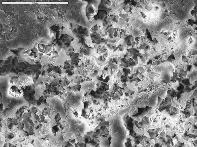
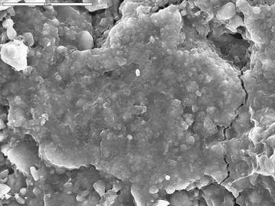
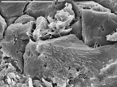
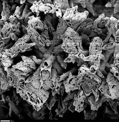
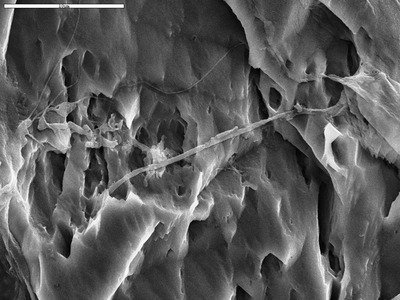
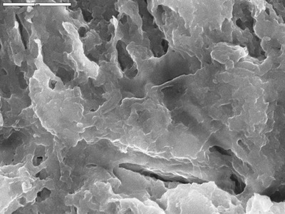
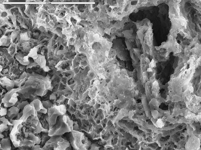
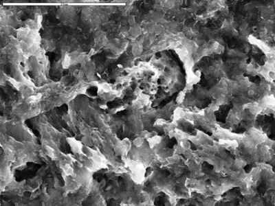
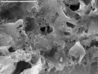
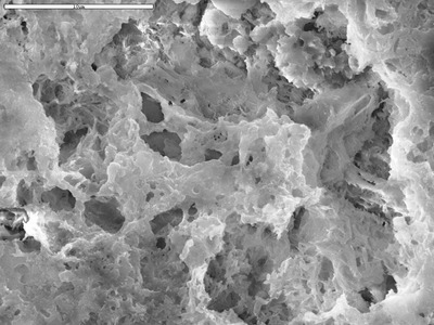
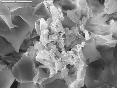
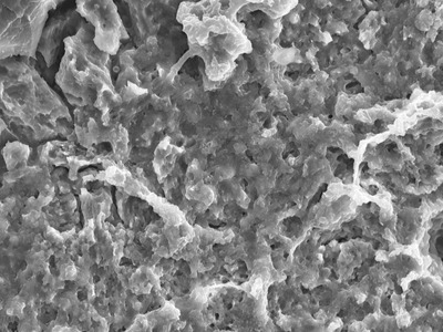
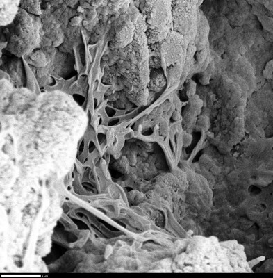
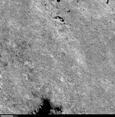
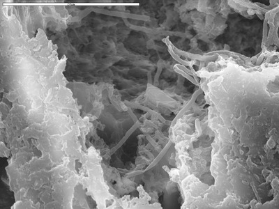
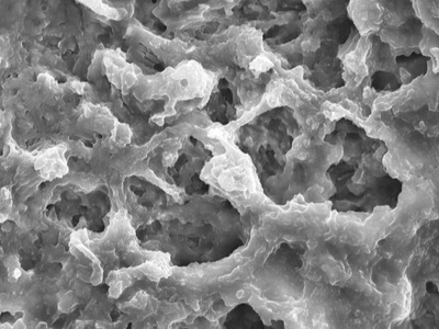
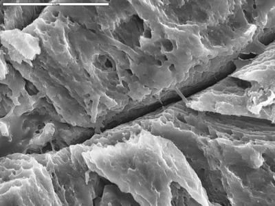
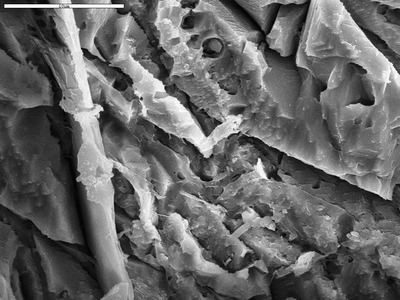
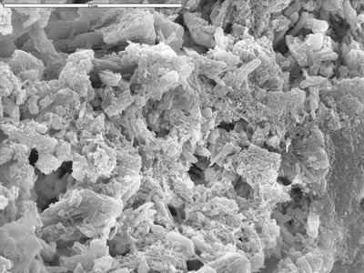
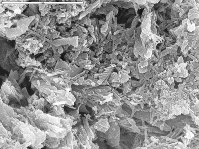
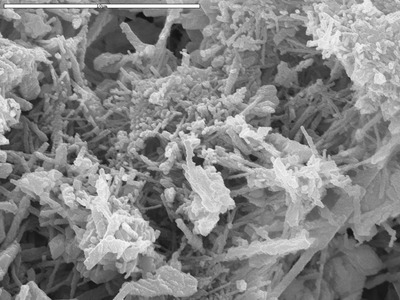
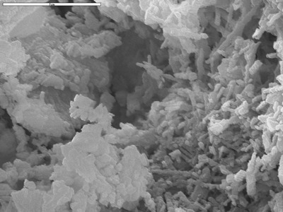
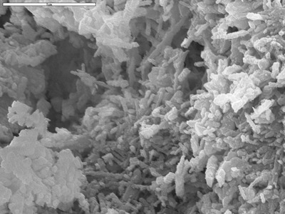
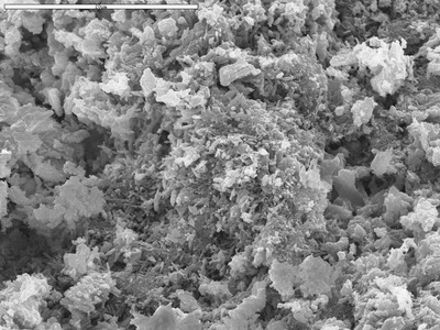
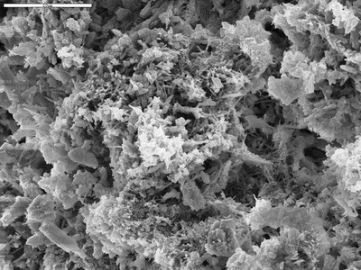
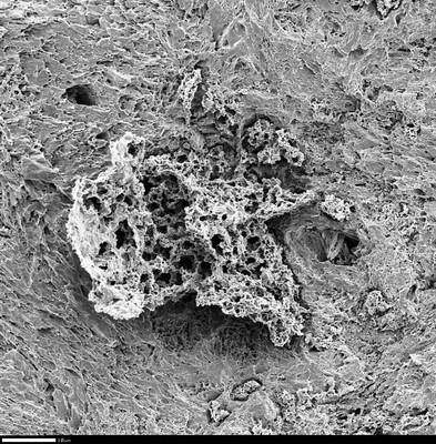
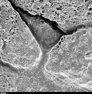
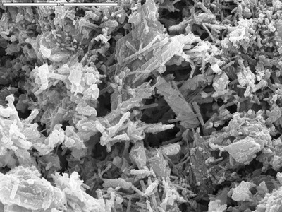
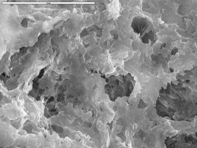
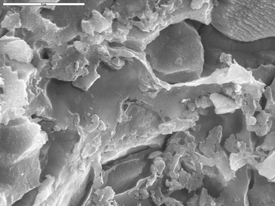
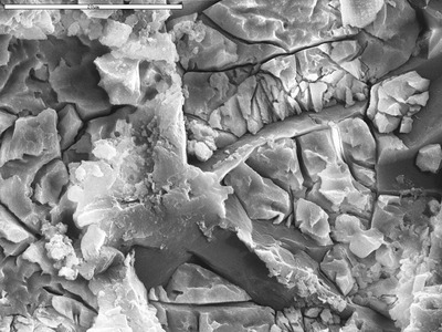
• Home
  • Search
  • Browse Collections
  • My Account
  • About
  • DC Network Digital Commons Network™
Skip to main content
UNM Digital Repository University of New Mexico UNM Digital Repository
  • Home
  • About
  • FAQ
  • My Account

Home > Lib, Mus, & Res Cts > Res Ctrs > Karst and Cave Studies > Pool Precipitates

Pool Precipitates

 
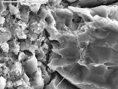
Scanning electron micrographs and spectra. All materials are copyrighted unless otherwise noted.
Printing is not supported at the primary Gallery Thumbnail page. Please first navigate to a specific Image before printing.

Follow

Switch View to List View Slideshow
 
  • Detail on filament with epoxy. by M. Spilde, D. Northup, and L. Melim

    Detail on filament with epoxy.

  • Detail on film below spheroid from L13-12 by M. Spilde

    Detail on film below spheroid from L13-12

  • Detail on film bridging between crystals by George Braybrook, Leslie Melim, and Brian Jones

    Detail on film bridging between crystals

  • Detail on film from qm8-27 by Michael Spilde

    Detail on film from qm8-27

  • Detail on large filament and film (to left)
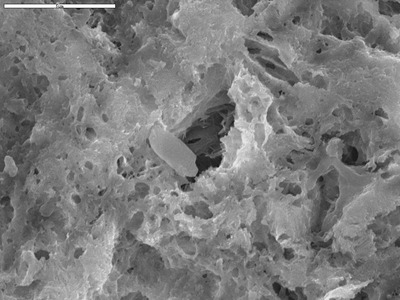
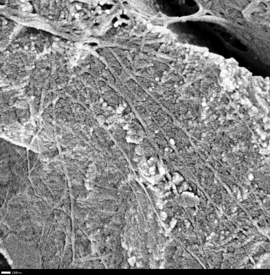
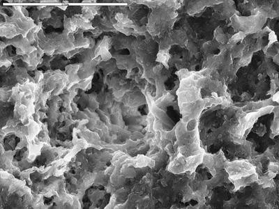
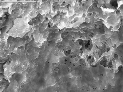
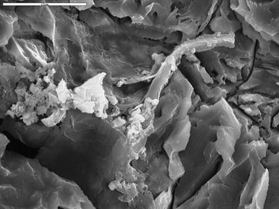
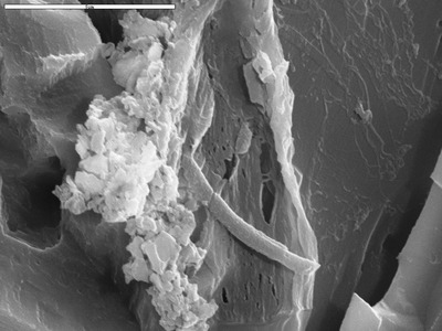
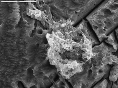
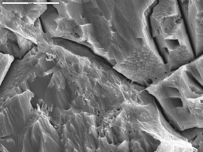
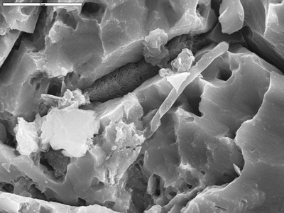
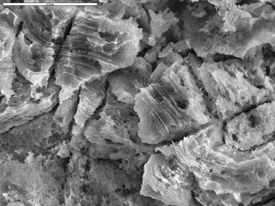
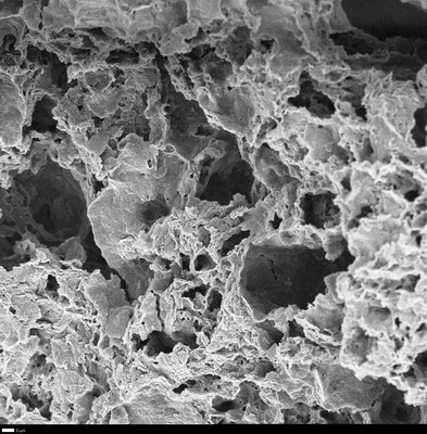
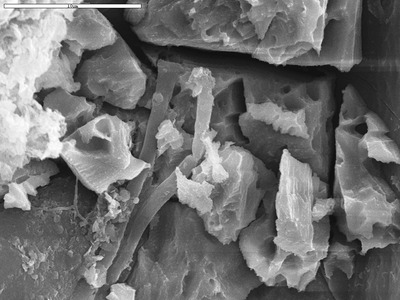
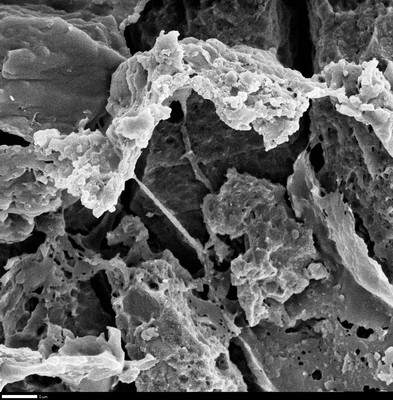
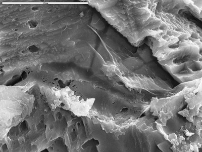
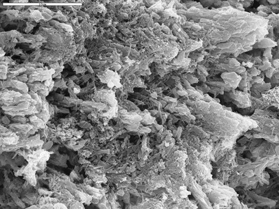
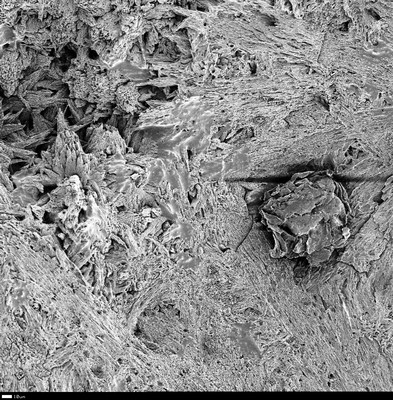
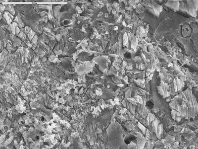
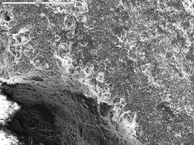
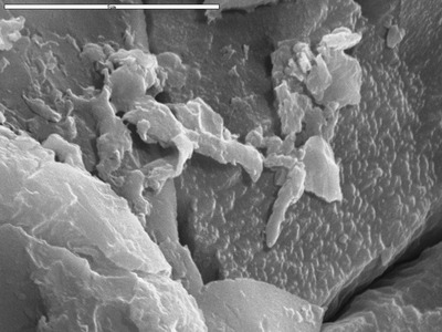
, flat area is epoxy, ropy area is not by Michael Spilde

    Detail on large filament and film (to left), flat area is epoxy, ropy area is not

  • Detail on layers in Q06-5a-20 with boundary to recrystallized area by M. Spilde, L. Melim, and D. Northup

    Detail on layers in Q06-5a-20 with boundary to recrystallized area

  • Detail on micrite texture. by D. Northup and L. Melim

    Detail on micrite texture.

  • Detail on needle etching out of large crystal by M. Spilde, D. Northup, and L. Melim

    Detail on needle etching out of large crystal

  • Detail on pair of filaments on crystal by M. Spilde and Leslie Melim

    Detail on pair of filaments on crystal

  • Detail on partially preserved reticulated filament by D. Northup, M. Splide, L. Melim, R. Liescheidt, and A. Kooser

    Detail on partially preserved reticulated filament

  • Detail on point of recrystallization cross-cutting the laminae by M. Spilde, D. Northup, and L. Melim

    Detail on point of recrystallization cross-cutting the laminae

  • Detail on point of recrystallization cross-cutting the laminae by M. Spilde, D. Northup, and L. Melim

    Detail on point of recrystallization cross-cutting the laminae

  • Detail on possible biofilm by D. Northup, M. Spilde, L. Melim, and R. Liescheidt

    Detail on possible biofilm

  • Detail on possible biofilm by M. Spilde, D. Northup, and L. Melim

    Detail on possible biofilm

  • Detail on possible biofilm by M. Spilde, D. Northup, and L. Melim

    Detail on possible biofilm

  • Detail on possible biofilm in J12B-29 by D. Northup, M. Spilde, L. Melim, and R. Liescheidt

    Detail on possible biofilm in J12B-29

  • Detail on possible biofilm in micrite by D. Northup and L. Melim

    Detail on possible biofilm in micrite

  • Detail on possible filament by D. Northup, M. Splide, L. Melim, R. Liescheidt, and A. Kooser

    Detail on possible filament

  • Detail on resistant feature sticking out of carbonate by D. Northup and L. Melim

    Detail on resistant feature sticking out of carbonate

  • Detail on reticulated filament and film by D. Northup and Leslie Melim

    Detail on reticulated filament and film

  • Detail on ropy filament attachment by George Braybrook, Leslie Melim, and Brian Jones

    Detail on ropy filament attachment

  • Detail on ropy film by D. Northup and L. Melim

    Detail on ropy film

  • Detail on round bodies and filament by George Braybrook, Leslie Melim, and Brian Jones

    Detail on round bodies and filament

  • Detail on round body/film by George Braybrook, Leslie Melim, and Brian Jones

    Detail on round body/film

  • Detail on smooth and hairy filaments. by George Braybrook, Leslie Melim, and Brian Jones

    Detail on smooth and hairy filaments.

  • Detail on smooth filament, about 0.5 micron in diameter by M. Spilde

    Detail on smooth filament, about 0.5 micron in diameter

  • Detail on specs etching out of calcite by M. Spilde

    Detail on specs etching out of calcite

  • Detail on spiral feature in lower right of L13-08 by M. Spilde

    Detail on spiral feature in lower right of L13-08

  • Detail on stretched film by D. Northup, M. Splide, L. Melim, R. Liescheidt, and A. Kooser

    Detail on stretched film

  • Detail on tiny star by George Braybrook, Leslie Melim, and Brian Jones

    Detail on tiny star

  • Detail on two reticulated filaments by D. Northup, M. Spilde, and L. Melim

    Detail on two reticulated filaments

  • Detail on very fine lamina showing etch. by M. Spilde, D. Northup, and L. Melim

    Detail on very fine lamina showing etch.

  • Detail on very long reticulated filament, chambers are larger than average by D. Northup, M. Splide, L. Melim, R. Liescheidt, and A. Kooser

    Detail on very long reticulated filament, chambers are larger than average

  • Detail, round body by George Braybrook, Leslie Melim, and Brian Jones

    Detail, round body

  • Detail, upper end of ropy film with round body by George Braybrook, Leslie Melim, and Brian Jones

    Detail, upper end of ropy film with round body

  • Detail view of large filament from qm8b-01, showing broken end and hollow inside by M. Spilde

    Detail view of large filament from qm8b-01, showing broken end and hollow inside

  • Dogtooth formations with broken crusts by George Braybrook, Leslie Melim, and Brian Jones

    Dogtooth formations with broken crusts

  • Edge of dense layer, shows fractured surface by George Braybrook, Leslie Melim, and Brian Jones

    Edge of dense layer, shows fractured surface

  • Edge of detrital core (NE half of image) 
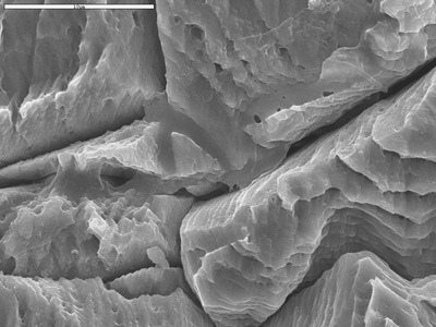
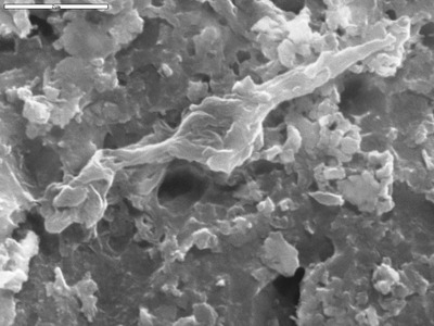
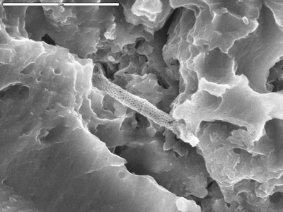
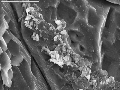
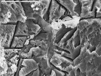
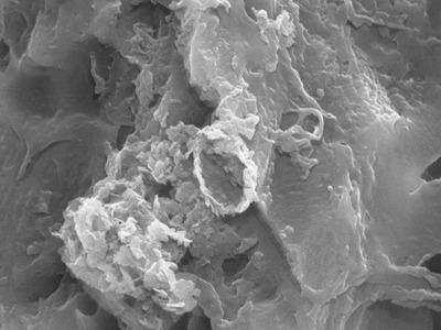
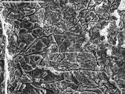
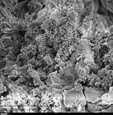
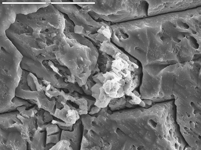
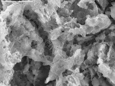
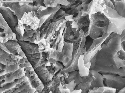
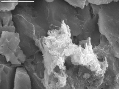
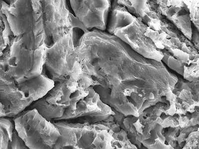
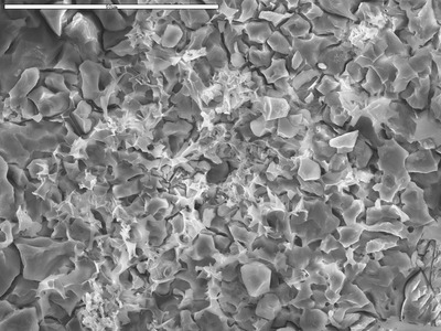
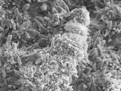
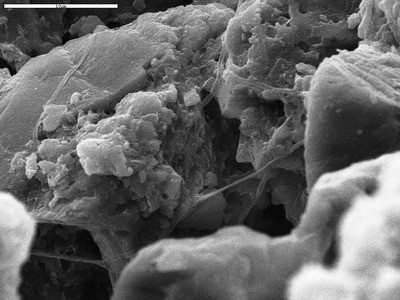
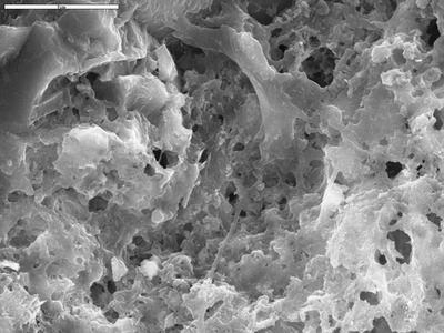
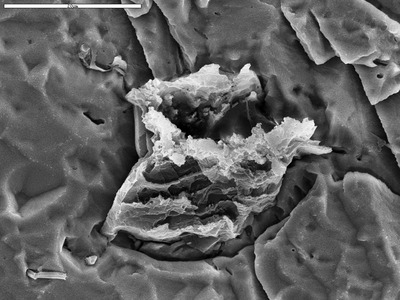
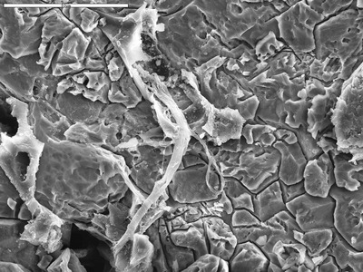
and inner layer of pearl by M. Spilde, D. Northup, and L. Melim

    Edge of detrital core (NE half of image) and inner layer of pearl

  • Edge of hydromagnestite by George Braybrook, Leslie Melim, and Brian Jones

    Edge of hydromagnestite

  • Edge of quartz grain with clays by D. Northup, M. Spilde, L. Melim, and R. Liescheidt

    Edge of quartz grain with clays

  • Edge of recrystallization point with epoxy ridge and possible area of biofilm by M. Spilde, D. Northup, and L. Melim

    Edge of recrystallization point with epoxy ridge and possible area of biofilm

  • Edge of resistant cracked film by George Braybrook, Leslie Melim, and Brian Jones

    Edge of resistant cracked film

  • EDS of epoxy bridge from hc2t_09.tif by M. Spilde, D. Northup, and L. Melim

    EDS of epoxy bridge from hc2t_09.tif

  • EDS on recrystallized area just next to laminae in Q06-5a-21. by M. Spilde, L. Melim, and D. Northup

    EDS on recrystallized area just next to laminae in Q06-5a-21.

  • EDS outside tube from q6-2b-14.tif showing epoxy by D. Northup and L. Melim

    EDS outside tube from q6-2b-14.tif showing epoxy

  • EDX behind globular grains from wen_11.tif by M. Spilde, D. Northup, and L. Melim

    EDX behind globular grains from wen_11.tif

  • EDX from 1048D_19.TIF by M. Spilde, D. Northup, and L. Melim

    EDX from 1048D_19.TIF

  • EDX from 1048E_05.TIF by M. Spilde, D. Northup, and L. Melim

    EDX from 1048E_05.TIF

  • EDX from HC1_06.tif by D. Northup and Leslie Melim

    EDX from HC1_06.tif

  • EDX from HC2b_10.tif showing clays by M. Spilde, D. Northup, and L. Melim

    EDX from HC2b_10.tif showing clays

  • EDX from W311B-10.TIF by M. Spilde, D. Northup, L. Melim, and R. Liescheidt

    EDX from W311B-10.TIF

  • EDX from wen_04.tif by M. Spilde, D. Northup, and L. Melim

    EDX from wen_04.tif

  • EDX lower right from HC1_40.tif by D. Northup and Leslie Melim

    EDX lower right from HC1_40.tif

  • EDX of area above cholla from d46-11 showing Carbon and iron oxides by M. Spilde

    EDX of area above cholla from d46-11 showing Carbon and iron oxides

  • EDX of area next to film from w312a_09.tif by M. Spilde, D. Northup, and L. Melim

    EDX of area next to film from w312a_09.tif

  • EDX of area of biofilm from d46-05 showing iron oxides by M. Spilde

    EDX of area of biofilm from d46-05 showing iron oxides

  • EDX of background crystals from wen_05.tif by M. Spilde, D. Northup, and L. Melim

    EDX of background crystals from wen_05.tif

  • EDX of background epoxy from d50-06 showing Cl peak by M. Spilde and Leslie Melim

    EDX of background epoxy from d50-06 showing Cl peak

  • EDX of background from c312a_02.tif by M. Spilde, D. Northup, and L. Melim

    EDX of background from c312a_02.tif

  • EDX of background from H292A_14.TIF, mostly likely epoxy by M. Spilde and L. Melim

    EDX of background from H292A_14.TIF, mostly likely epoxy

  • EDX of background from HC1_37.tif showing clay by D. Northup and Leslie Melim

    EDX of background from HC1_37.tif showing clay

  • EDX of background from HC3a_02.tif by M. Spilde, D. Northup, and L. Melim

    EDX of background from HC3a_02.tif

  • EDX of background from HC4_23.tif by M. Spilde and Leslie Melim

    EDX of background from HC4_23.tif

  • EDX of background from wen_03 EDS.tif by M. Spilde, D. Northup, and L. Melim

    EDX of background from wen_03 EDS.tif

  • EDX of background near filaments from 1022_12.TIF, showing possible biofilm by M. Spilde, D. Northup, and L. Melim

    EDX of background near filaments from 1022_12.TIF, showing possible biofilm

  • EDX of background near filaments from HC1_19.tif by D. Northup and Leslie Melim

    EDX of background near filaments from HC1_19.tif

  • EDX of barite patch from C852B_03.tif by M. Spilde, D. Northup, and L. Melim

    EDX of barite patch from C852B_03.tif

  • EDX of biofilm fold in upper center of d46-05 showing calcium by M. Spilde

    EDX of biofilm fold in upper center of d46-05 showing calcium

  • EDX of bright area in HC2b_03.tif by M. Spilde, D. Northup, and L. Melim

    EDX of bright area in HC2b_03.tif

  • EDX of bright spot from tp1-06.tif by M. Spilde, L. Melim, and D. Northup

    EDX of bright spot from tp1-06.tif

  • EDX of bright spots from BSE HC3a_06.tif by M. Spilde, D. Northup, and L. Melim

    EDX of bright spots from BSE HC3a_06.tif

  • EDX of calcite crystals from hc3d_23.tif by M. Spilde and Leslie Melim

    EDX of calcite crystals from hc3d_23.tif

  • EDX of center area of d46-22 showing silica by M. Spilde

    EDX of center area of d46-22 showing silica

  • EDX of center from 1048D_10.TIF by M. Spilde, D. Northup, and L. Melim

    EDX of center from 1048D_10.TIF

  • EDX of center spheroids from HC1_44.tif by D. Northup and Leslie Melim

    EDX of center spheroids from HC1_44.tif

  • EDX of cholla from d46-11 showing carbon by M. Spilde

    EDX of cholla from d46-11 showing carbon

  • EDX of clay from 1048D_07.TIF by M. Spilde, D. Northup, and L. Melim

    EDX of clay from 1048D_07.TIF

  • EDX of clay from H302A_01.TIF by M. Spilde, D. Northup, and L. Melim

    EDX of clay from H302A_01.TIF

  • EDX of clay from hc2t_15.tif by M. Spilde, D. Northup, and L. Melim

    EDX of clay from hc2t_15.tif

  • EDX of clay from HC3d_19.tif by M. Spilde and Leslie Melim

    EDX of clay from HC3d_19.tif

  • EDX of clay from HC3d_19.tif by M. Spilde and Leslie Melim

    EDX of clay from HC3d_19.tif

  • EDX of clay from hc3d_23.tif by M. Spilde and Leslie Melim

    EDX of clay from hc3d_23.tif

  • EDX of clay from J12B-10.tif by D. Northup, M. Spilde, L. Melim, and R. Liescheidt

    EDX of clay from J12B-10.tif

  • EDX of clay from qm8-06 by M. Spilde

    EDX of clay from qm8-06

  • EDX of clay in boundary layer by M. Spilde, D. Northup, and L. Melim

    EDX of clay in boundary layer

  • EDX of clay in HC1_37.tif by D. Northup and Leslie Melim

    EDX of clay in HC1_37.tif

  • EDX of clay layer by M. Spilde, D. Northup, and L. Melim

    EDX of clay layer

  • EDX of clay on end of upper filament from d50-03 showing clay by M. Spilde and Leslie Melim

    EDX of clay on end of upper filament from d50-03 showing clay

  • EDX of clay over reticulated filament from qm8-25, showing carbon by M. Spilde

    EDX of clay over reticulated filament from qm8-25, showing carbon

  • EDX of clay patch from LHT2_10.tif by M. Spilde, D. Northup, and L. Melim

    EDX of clay patch from LHT2_10.tif

  • EDX of clays from c851a_06.tif by M. Spilde, D. Northup, and L. Melim

    EDX of clays from c851a_06.tif

  • EDX of clays on calcite from LHT2_02.tif by M. Spilde, D. Northup, and L. Melim

    EDX of clays on calcite from LHT2_02.tif

  • EDX of clays on calcite from LHT2_02.tif by M. Spilde, D. Northup, and L. Melim

    EDX of clays on calcite from LHT2_02.tif

  • EDX of coated filament from 1024_19.TIF by D. Northup and Leslie Melim

    EDX of coated filament from 1024_19.TIF

  • EDX of crust from 1048D_27.TIF by M. Spilde, D. Northup, and L. Melim

    EDX of crust from 1048D_27.TIF

  • EDX of crust from 1048D_27.TIF by M. Spilde, D. Northup, and L. Melim

    EDX of crust from 1048D_27.TIF

  • EDX of crust from 1048D_27.TIF by M. Spilde, D. Northup, and L. Melim

    EDX of crust from 1048D_27.TIF

  • EDX of crystal from tp1-16.tif by M. Spilde, L. Melim, and D. Northup

    EDX of crystal from tp1-16.tif

  • EDX of crystals from L13-19 showing iron and calcium by M. Spilde

    EDX of crystals from L13-19 showing iron and calcium


Page 6 of 16

  • 3
  • 4
  • 5
  • 6
  • 7
  • 8
  • 9


Search

Advanced Search

  • Notify me via email or RSS

Browse

  • Collections
  • Disciplines
  • Authors

Author Corner

  • Author FAQ

Gallery Locations

  • View gallery on map
  • View gallery in Google Earth
 
Elsevier - Digital Commons

Home | About | FAQ | My Account | Accessibility Statement

Privacy Copyright