


Document Type
Article
Publication Date
1-1-2021
Abstract
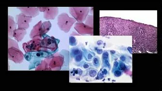
Florid mesothelial hyperplasia typically occurs in the pelvis, abdomen, or chest associated with an underlying neoplastic or inflammatory process. These lesions are of clinical significance because they can mimic a neoplasm. Early reports were published in the 1970s, but only a few case series of such lesions have been published in the gynecologic pathology literature. Here, we report a case of florid mesothelial hyperplasia with an infiltrative growth pattern, mimicking an invasive carcinoma. The lesion was associated with endometriosis forming a mass lesion in the abdominal wall. Histologically, tubular arrangements and nests of mesothelial cells, some with artifactual slit-like spaces, formed a stellate lesion adjacent to endometrial glands and stroma. Cytologic atypia was mild and reactive, and positive immunostaining for calretinin, WT-1, and cytokeratin 5 identified the lesion as mesothelial and benign. We describe in detail the histologic findings in this case and review the pertinent literature. We discuss the clinically importance of this diagnostic pitfall and the path to arriving at the correct diagnosis.
Recommended Citation
Fischer EG, Agarwal S. Florid Mesothelial Hyperplasia Associated with Abdominal Wall Endometriosis Mimicking Invasive Carcinoma. Case Rep Pathol. 2021 Nov 28;2021:3439700. doi: 10.1155/2021/3439700. PMID: 34877024; PMCID: PMC8645372.