


Document Type
Article
Publication Date
10-1-2019
Abstract
occidioidomycosis is a fungal infection which is prevalent in the southwestern region of the United States.1 Clinical presentation of pulmonary coccidioidomycosis is nonspecific, and the disease can manifest as subclinical infection, upper respiratory viral-like illness, and rarely severe respiratory failure requiring intensive care admission. Radiologically the disease can present with pulmonary infiltrates, nodules or cavitary lesions which can mimic other infectious, autoimmune, and neoplastic disorders.2 The diagnosis is typically made using serologic testing.3 The most commonly performed serologic methods are enzyme-linked immunoassay (EIA), qualitative immunodiffusion testing for immunoglobulin M and G antibodies and quantitative complement fixation.3 EIA testing has a high sensitivity, but low specificity, so a positive EIA test should be confirmed with either immunodiffusion fixation or complement fixation testing which has lower sensitivity but higher specificity and positive predictive value.3
It is important to note that it can take from 7 days to several weeks for antibodies to become detectable, so negative serologic testing does not eliminate the possibility of disease, especially in the early course or in immunocompromised individuals. Sputum culture lacks sensitivity but if positive is specific for pulmonary coccidioidomycosis; of note, laboratory personnel must be notified if coccidioidomycosis is suspected given the high contagious risk of the organism.4
Often, bronchoscopy with bronchoalveolar lavage (BAL) is performed in cases with clinically suspected coccidioidomycosis, despite negative serology. The sensitivity of bronchoscopy with BAL is higher in patients with pulmonary infiltrates, endoluminal infection and cavitary lesions.5,6 DiTomasso et al7 compared the test characteristics of BAL with or without bronchial washings to transbronchial biopsy. The sensitivity of BAL and bronchial washings ranged from 31% to 42% compared with 100% of transbronchial needle aspiration (TBNA). Of note, TBNA was performed only in select patients which may imply a selection bias.
Shah et al8 retrospectively reviewed 13 cases of pulmonary coccidioidomycosis diagnosed with endoscopic ultrasound FNA and EBUS-TBNA of the mediastinal and hilar lymph node. Bronchoscopy with BAL was positive in 38% of patients compared with 82% of EBUS-TBNA and 55% of endoscopic ultrasound FNA.
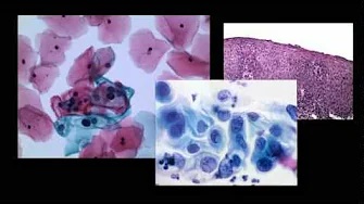
To the best of our knowledge, we report a first documented case of pulmonary coccidioidomycosis which was diagnosed with EBUS-FNA biopsy of the 17-mm solid pulmonary paratracheal nodule published in a peer-reviewed academic journal. Of note, Hyman et al9 presented a poster at American College of Chest Physician meeting in 2018 of a patient with endobronchial lesion who was diagnosed with coccidioidomycosis via EBUS and TBNA. However, our patient had negative coccidioidomycosis serology. Of note, many patients with residual and asymptomatic nodular pulmonary coccidioidomycosis have negative serology.10 We propose that EBUS-FNA biopsy as an effective method to sample pulmonary parenchymal nodules to diagnose pulmonary coccidioidomycosis and exclude other pathologies.
Recommended Citation
Mirrakhimov AE, Hnatiuk O, Grant T, Martin DR, Saeed AI. Pulmonary Coccidioidomycosis Diagnosed by Endobronchial Ultrasound With Fine Needle Aspiration Biopsy of a Paratracheal Pulmonary Nodule. J Bronchology Interv Pulmonol. 2019 Oct;26(4):e63-e65. doi: 10.1097/LBR.0000000000000616. PMID: 31569107.