


Document Type
Article
Publication Date
1-1-2020
Abstract
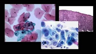
A small subepithelial esophageal lesion was seen 21 cm from the incisors in an otherwise unremarkable examination. Subsequent upper-EUS examination revealed a 14 mm by 21 mm well-circumscribed, hypoechoic lesion originating from the muscularis propria (layer 4). The initial EUS impression was suggestive of a GI stromal tumor, however, examination of EUS-guided fine-needle biopsy specimens showed a haphazard arrangement of spindle cells in a fibrotic stoma.
Recommended Citation
Gulati R, Hanson JA, Parasher G, Castresana D. Getting the gist of a schwannoma. Gastrointest Endosc. 2020 Jan;91(1):191-192. doi: 10.1016/j.gie.2019.07.027. Epub 2019 Aug 2. PMID: 31381899.
COinS