


Document Type
Article
Publication Date
12-1-2019
Abstract
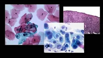
Case 1 A previously healthy 16-year-old man was initially evaluated in the emergency department for traumatic injuries following a high-impact motor vehicle accident. He was a bodybuilder who used anabolic steroids. He denied abdominal or back pain. His physical examination was signifcant for a palpable liver edge. Routine laboratory tests including liver function tests were unremarkable. A computed tomographic (CT) scan showed an unsuspected mass in the right lobe of the liver. A subsequent triple-phase magnetic resonance imaging (MRI) scan of the abdomen showed a 15.1 × 15.0 × 10.8 cm mass in the right hepatic lobe that exhibited heterogeneous arterial and thin capsular enhancement, concerning for a hepatic neoplasm (Fig. 1). He underwent partial hepatectomy; the resected surgical specimen showed a well-differentiated hepatocellular carcinoma that on immunohistochemical staining (Fig. 2a) showed β-catenin activation via aberrant nuclear expression. The tumor was confned to the liver with no vascular invasion or regional lymph node involvement. It was predominantly composed of bland hepatocytes, though there were areas of scattered cytological atypia. Moreover, the architecture was difusely pseudoglandular (Fig. 2b) with reticulin staining showing multifocal loss of the normal peri-cellular reticulin framework (Fig. 2c). These latter features were considered diagnostic of a well-diferentiated hepatocellular carcinoma (HCC) as opposed to a β-catenin-activated hepatic adenoma (HCA). The patient and his guardian decided against undergoing chemotherapy. The patient is being followed up by the pediatric oncology service, to monitor for possible tumor progression, with periodic surveillance by imaging with MRI. C
Recommended Citation
Ling C, Khalid S, Martin D, Hanson J, Castresana D, McCarthy D. HCCs and HCAs in Non-cirrhotic Patients: What You See May Not Be Enough. Dig Dis Sci. 2019 Dec;64(12):3440-3445. doi: 10.1007/s10620-019-05920-z. PMID: 31673903.