Authors

Alexander D. Borowsky, Department of Pathology and Laboratory Medicine, University of California, Davis, Sacramento
Eric F. Glassy, The Affiliated Pathologists Medical Group, Rancho Dominguez, California
William Dean Wallace, UCLA Pathology & Lab Medicine, University of California, Los Angeles
Nathash S. Kallichanda, The Affiliated Pathologists Medical Group, Rancho Dominguez, California
Cynthia A. Behling, The Pacific Rim Pathology Lab and Sharp Healthcare, San Diego, California
Dylan V. Miller, Intermountain Central Laboratory, Salt Lake City, Utah
Hemlata N. Oswal, The Pathology Department, Lucent Pathology Partners Mercy San Juan Hospital, Carmichael, California
Richard M. Feddersen, The Histology Lab, TriCore Reference Laboratories, Albuquerque, New Mexico
Omid R M. Bakhtar, Scripps Clinic Torrey Pines, La Jolla, California
Arturo E. Mendoza, The Pacific Rim Pathology Lab and Sharp Healthcare, San Diego, California
Daniel P. Molden, The Pacific Rim Pathology Lab and Sharp Healthcare, San Diego, California
Helene L. Saffer, The Pacific Rim Pathology Lab and Sharp Healthcare, San Diego, California
Christopher R. Wixom, The Pacific Rim Pathology Lab and Sharp Healthcare, San Diego, California
James E. Albro, Intermountain Central Laboratory, Salt Lake City, Utah
Melissa H. Cessna, Intermountain Central Laboratory, Salt Lake City, Utah
Brian J. Hall, Intermountain Central Laboratory, Salt Lake City, Utah
Isaac E. Lloyd, Intermountain Central Laboratory, Salt Lake City, Utah
John W. Bishop, Department of Pathology and Laboratory Medicine, University of California, Davis, Sacramento
Morgan A. Darrow, Department of Pathology and Laboratory Medicine, University of California, Davis, Sacramento
Dorina Gui, Department of Pathology and Laboratory Medicine, University of California, Davis, Sacramento
Kuang-Yu Jen, Department of Pathology and Laboratory Medicine, University of California, Davis, Sacramento
Julie Ann S. Walby, Department of Pathology and Laboratory Medicine, University of California, Davis, Sacramento
Stephen M. Bauer, The Pathology Department, Lucent Pathology Partners Mercy San Juan Hospital, Carmichael, California
Daniel A. Cortez, The Pathology Department, Lucent Pathology Partners Mercy San Juan Hospital, Carmichael, California
Pranav Gandhi, Scripps Clinic Torrey Pines, La Jolla, California
Melissa M. Rodgers, The Pathology Department, Lucent Pathology Partners Mercy San Juan Hospital, Carmichael, California
Rafael A. Rodriguez, The Pathology Department, Lucent Pathology Partners Mercy San Juan Hospital, Carmichael, California
David R. Martin, The Histology Lab, TriCore Reference Laboratories, Albuquerque, New Mexico
Thomas G. McConnell, The Histology Lab, TriCore Reference Laboratories, Albuquerque, New Mexico
Samuel J. Reynolds, The Histology Lab, TriCore Reference Laboratories, Albuquerque, New Mexico
James H. Spigel, The Histology Lab, TriCore Reference Laboratories, Albuquerque, New Mexico
Shelly A. Stepenaskie, The Histology Lab, TriCore Reference Laboratories, Albuquerque, New Mexico
Elena Viktorova, Beckman Coulter, Inc., Miami, Florida
Robert Magari, Beckman Coulter, Inc., Miami, Florida
Keith A. Wharton, Leica Biosystems Imaging, Inc., Danvers, Massachusetts
Jinsong Qiu, Leica Biosystems, Vista, California
Thomas W. Bauer, The Department of Pathology and Laboratory Medicine, Hospital for Special Surgery, Weill Cornell Medical College, New York, New Yor

Document Type

Article

Publication Date

10-1-2020

Abstract

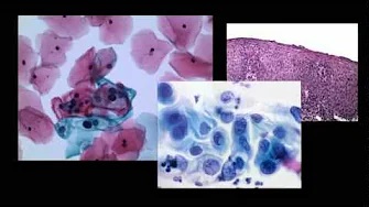
CONTEXT.—: The adoption of digital capture of pathology slides as whole slide images (WSI) for educational and research applications has proven utility.

OBJECTIVE.—: To compare pathologists' primary diagnoses derived from WSI versus the standard microscope. Because WSIs differ in format and method of observation compared with the current standard glass slide microscopy, this study is critical to potential clinical adoption of digital pathology.

DESIGN.—: The study enrolled a total of 2045 cases enriched for more difficult diagnostic categories and represented as 5849 slides were curated and provided for diagnosis by a team of 19 reading pathologists separately as WSI or as glass slides viewed by light microscope. Cases were reviewed by each pathologist in both modalities in randomized order with a minimum 31-day washout between modality reads for each case. Each diagnosis was compared with the original clinical reference diagnosis by an independent central adjudication review.

RESULTS.—: The overall major discrepancy rates were 3.64% for WSI review and 3.20% for manual slide review diagnosis methods, a difference of 0.44% (95% CI, -0.15 to 1.03). The time to review a case averaged 5.20 minutes for WSI and 4.95 minutes for glass slides. There was no specific subset of diagnostic category that showed higher rates of modality-specific discrepancy, though some categories showed greater discrepancy than others in both modalities.

CONCLUSIONS.—: WSIs are noninferior to traditional glass slides for primary diagnosis in anatomic pathology.

Share

COinS